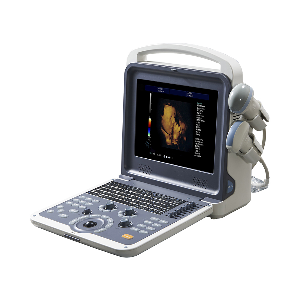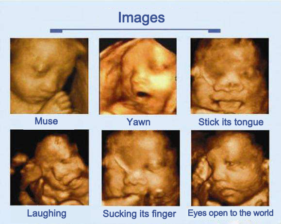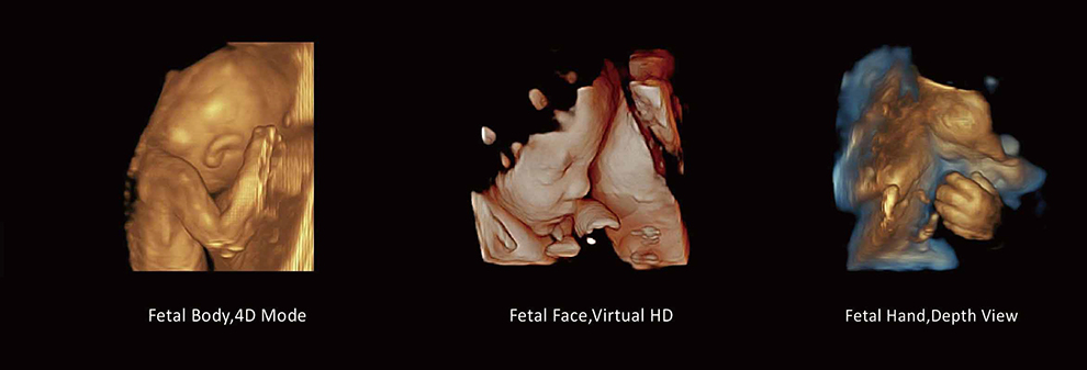
Four-dimensional color ultrasonic diagnostic apparatus is the world's most advanced Color Doppler Ultrasound equipment. It can show a real-time animated image of your unborn baby, or a real-time moving image of other organs in the body.
"4D" is the abbreviation of "four dimensions". The fourth dimension refers to the time of this vector, so it is also called real-time three-dimensional. For ultrasound field, 4D ultrasound technology is a revolutionary breakthrough. 4D ultrasound technology is the use of 3D ultrasound images with time dimension parameters, the revolutionary technology to real-time access to three-dimensional images, beyond the traditional ultrasound restrictions. It is used in many fields including abdomen, blood vessels, small organs, obstetrics, gynecology, urology, neonatal and pediatric.
4D color Doppler ultrasound is not only feel the baby's breathing and exercise, but can witness their every move and well-behaved look. More importantly, the four-dimensional color Doppler ultrasound can be multi-directional, multi-angle observation of intrauterine fetal growth and development, for the early diagnosis of fetal congenital malformations and congenital heart disease to provide accurate scientific basis.
Color Doppler Ultrasound, 3D (three-dimensional) color Doppler ultrasound, 4D (four-dimensional) color Doppler ultrasound. Color of their images are actually B&W (black and white), and 3D Color Doppler Ultrasound, 4D Color Doppler Ultrasound made three-dimensional, four-dimensional images, and then add color to the image to mark the information, images of Color Doppler Ultrasound is not colorful actually. We call it color Doppler ultrasound, because it will use color marked heart, blood flow and other informations. Color Doppler Ultrasound higher resolution than the general Black and white ultrasound. When doctors need more detailed informations to diagnose, more doctors are willing to diagnose through the color Doppler ultrasound. And as blood flow is marked with color, when the umbilical cord around the neck , you will see the baby's neck was U-shaped or W-shaped blood flow, Whether the umbilical cord is around the neck or not, it is clear for us.
3D color Doppler ultrasound and 4D color Doppler ultrasound is the difference lies in a "time dimension", that is, 3D color Doppler ultrasound is a “photo”, 4D color Doppler ultrasound is a “video”, you can let pregnant mother see a series of fetal movements.

The 4D color Doppler ultrasound is dynamic, 3D color Doppler ultrasound is static, so the 4D color Doppler ultrasound will be more clear.
3D color Doppler ultrasound is only a certain time point on the photo, the 4D color Doppler ultrasound can be made it as a video , can burn a disc. 3D color Doppler ultrasound and HD 4D color Doppler ultrasound has the same funtion of fetal anomaly diagnosis, but HD 4D color Doppler ultrasound more accurate.
Four-dimensional color Doppler ultrasound diagnosis of fetal mainly include the following aspects:
Fetal facial deformities: such as cleft lip and palate and the like.
2. Nervous System: no brain child, hydrocephalus, microcephaly, spina bifida and meningocele.
3. Digestive system: umbilical intestinal bulge, visceral valgus, intestinal atresia and megacolon and so on.
4. Urinary System: hydronephrosis, polycystic kidney disease and giant bladder, urethral obstruction.
5. Other deformities: short limb deformity, conjoined deformity, cleft lip, four heart chamber.
6. Amniotic fluid too much, too little.
Use of 4d color Doppler ultrasound
·Determine fetal age
·Evaluate multiple births or high-risk pregnancy
·Analyze the development of the fetus
·Detection of placental abnormalities
·Detection of ectopic pregnancy and other abnormal pregnancy
·Detection of uterine structural abnormalities
Tips
About obstetric examination
Any pregnancy weeks can do 4D color Doppler ultrasound examination, but need to emphasize that, for safety reasons, it is recommended to do 4D color Doppler ultrasound examination after 16-week.
After 16 weeks of pregnancy, fetus limbs and the main organs have all developed, the best time to check for 16 to 24 weeks of pregnancy, and amniotic fluid more suitable for fetal malformations.

