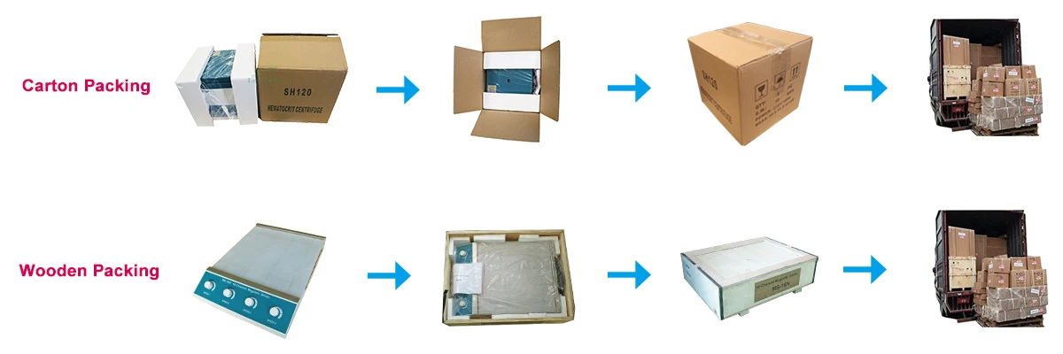
We ship goods by UPS/DHL/FEDEX/TNT express takes 3~ 5 days to arrive.

LTUB15 Trolley color doppler ultrasound scanner
1. The host system
1.1 Full-digital color Doppler ultrasonic diagnostic system
1.2 Ultrasound host operating system: Windows XP operating system
1.3 Monitor: ≥ 17-inch high-definition professional medical LED displays, with a 7-inch widescreen touch-navigation system
1.4 Probe connector (all effectively activate) ≥ 3
1.5 2D gray-scale imaging unit
1.6 The PW pulse wave Doppler
1.7 Doppler energy and directional energy diagram
1.8 Color Doppler ultrasound diagnostic unit
1.9 The THI tissue harmonic Imaging
Full (Wide) scene imaging
Extend the (extended) pulse imaging (EPI)
Real-time 3D/4D synchronization imaging
Trapezoidal extended imaging
Anatomical M-mode imaging
Elasticity imaging (ELASTOGRAPH)
1.10 Inverting pulse harmonic imaging
1.11 A key optimization (optimize 2D gray-scale images and PW images)
1.12 Multi lingual user interface optional, Chinese/English operation interface, support for Chinese/English input.
1.13 Real-time synchronization unit: 2D, color Doppler, spectral Doppler / continuous CWD at the same time the same screen real-time display.
1.14 Professional package: abdomen, obstetrics and gynecology, small parts, urology, orthopedic surgery, cardiac
1.15 Measurement: distance, area, angle, time, slope, heart rate, speed, acceleration, spectrum tracing, resistance index and pulsate index
1.16 Gain: B / PW / CD can be separately adjusted
1.17 Movie playback ≥ 1000 frame
2. Probe configuration and specifications:
2.1 Configuration: convex array probe, the high-frequency linear array probe, transvaginal probe
2.2 convex array probe :2.5-5 .0 MHz center frequency: ≥ 3, visual adjustable; with harmonic imaging capabilities, the harmonic frequency of ≥ 2, visual adjustable
2.3 linear probe :6.0-10 .0 MHz center frequency: ≥ 3, visual adjustable; with harmonic imaging capabilities, the harmonic frequency≥ 2, visual adjustable
2.4 Transvaginal probe : 5.0-8 .0 MHz (scan angle ≥ 130°)
2.5 Variable frequency phased array cardiac probe: the frequency range 2 - 4MHz, 3 frequency bandwidth is optional; probe scanning angle:10° to 85° .
3. 2D imaging modes:
3.1 4B imaging mode
3.2 B-type images in real time and freeze the zoom magnification of ≥ 8, 10 or more adjustable
3.3 Grayscale: 256
3.4 Position chart: ≥ 60
3.5 Dynamic range: 0-150db, visual adjustable 10 level and above
3.6 The probing depth: ≥ 360mm
3.7 Resolution: lateral ≤ 2mm; vertical ≤ 1mm
3.8 Focus: B mode, the focus position and focus distance is adjustable
3.9 Ultrasonic power output: ≥ 10 adjustable
3.10 Preconditions: ≥ 10 species, the user can customize the conditions
3.11 Pseudo-color display: ≥ 7 species
3.12 Convex array probe: 450px depth, full aspects, 2D frame rate≥ 30 frames / sec.
3.13 Linear array probe beam deflection
3.14 Digital beam formation: continuous dynamic focusing, variable aperture and dynamic change trajectory
3.15 B, 2B, 4B display modes
3.16 TGC segment gain adjustment ≥ 8 segments
4. M model
4.1 B / M to a variety of layout adjustable
4.2 M scanning speed is adjustable
5. Color Doppler:
5.1 Color gain and PRF multi-level adjustable
5.2 Visual Color frequency adjustable: ≥ 2
5.3 Sampling frame: the size and position adjustable
5.4 Color Atlas: ≥ 7
5.5 Color afterglow: ≥ 4
5.8 Color baseline adjustable
Color Doppler energy diagram:
6.1 The direction of the energy diagram
6.2 Energy in gain: Adjustable
6.3 The energy diagram
7. Doppler spectrum:
7.1 Doppler spectrum of the emission frequency can be multi-file variable frequency
7.2 The measurement speed: the minimum detectable speed of 1mm / s maximum speed measurement ≥ 25m / s,
7.3 Sample volume size: 1mm-15mm, adjustable
7.4 Sampling point of correction: -70 ° to 70°
7.5 Spectrum Gain: adjustable;
7.6 Spectrum Automatic tracing and automatic measurement functions, including automatic envelope, manual envelope, fast measurement
7.7 Tracings range to support the baseline above and below the baseline, and all tracings in three ways
7.8 Baseline: Zero moving grading adjustable ≥ 8 levels
7.9 Scan speed: ≥ 3 adjustable
7.10 Wall Filtering: ≥ 5-level adjustable
7.11 Spectrum volume size is adjustable
7.12 Spectrum envelope automatically in real time measurement and calculation
8. Iimage-text management system:
8.1 Built-in storage unit: hard drive storage capacity ≥ 160G;
8.2 Built-in ultrasound image archiving and management functionality: You can edit the diagnostic report , the ultrasound diagnostic images can be embedded in the report and print directly
Q:What's the minimum order quantity (MOQ)?
A:For most of our medical products, even order for only one unit is warmly welcomed.
Q: Can you do OEM/ private label?
A: Of course we can do OEM/private label for you.
Q: What's your delivery time?
A: Generally it is 10 days if the goods are in stock.
or it is 15-20 days if the goods are not in stock, it is according to quantity.
Q: How to ship the order?
A: Please inform us your instruction, by sea, by air or by express, any way is ok for us. We have very professional forwarder to provide the best shipping cost, service and guarantee.
Q: What is your terms of payment ?
A: We accept T/T ,LC,Western Union ,Paypal and more.Please suggest your preferred payment method.